Examination is performed by an experienced dermatologist
Catching malignant growth at an early stage
Possibility of monitoring the development of skin changes
Description of the Examination
Diagnosis of Skin Lesions
Although our understanding of the causes of cancer continues to grow, modern medicine cannot always prevent these diseases. That is precisely why screening tests (known as secondary prevention) are available. These tests are intended for virtually everyone, including healthy individuals and those at low risk.
What Is Dermoscopy?
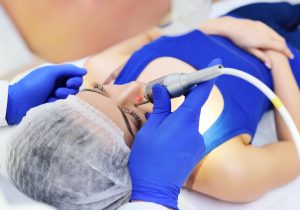
The goal of the examination is to detect malignant tumors in their early stages, when treatment is usually relatively simple. Mole screening also aims to identify precancerous moles and prevent the development of skin cancer. At our clinic, we use high-quality handheld dermatoscopes to examine moles, which enable us to provide fast and reliable diagnoses.
At our clinic, we also offer examinations using a digital dermatoscope. Unlike a handheld device, it takes images of moles, which it stores, allowing for comparison of changes over time. However, ordinary, benign moles do not require long-term monitoring with a digital dermatoscope. It is particularly suitable for patients who have multiple so-called dysplastic nevi on their bodies.
These moles show changes over time, but are not yet dangerous. On the other hand, however, they are associated with a higher risk of developing cancer. The World Health Organization (WHO) does not recommend removing these moles immediately, but rather monitoring them for now. In practice, patients are first examined with a manual dermatoscope, and if our experienced dermatologist deems it necessary, a digital examination follows.
How We Grade Each Symbol
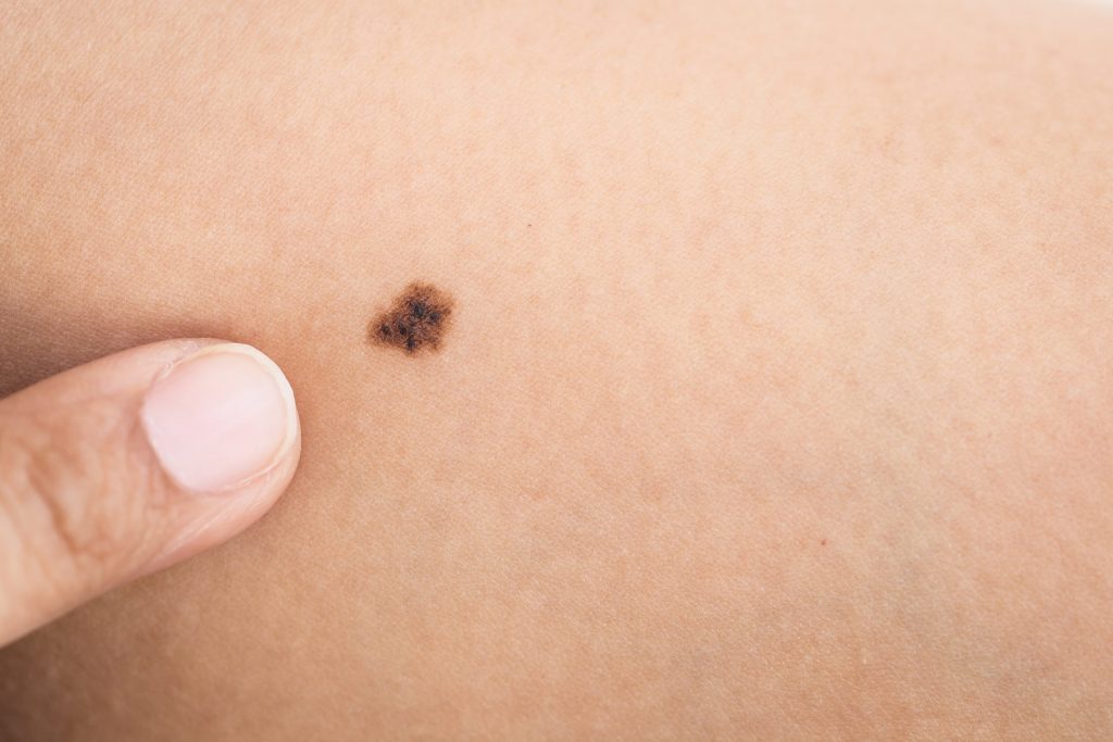
We thoroughly examine every mole on our clients’ bodies and evaluate it based on several criteria. Using these criteria, we can determine whether a mole is safe, requires monitoring, or should be removed. The evaluation criteria are as follows:
A (asymmetry) – the overall regularity of the sign.
B (border) – the border of the mark may be sharp, blurred, or have irregular projections.
C (color) – The mark has a shade of red or brown, or may contain various shades of these colors.
D (diameter/dimension) – small characters have a diameter of up to 6 mm, and large characters have a diameter greater than 6 mm.
E (evolution) – evolution over time based on changes in the parameters listed above.
When should you go for a checkup?
If the mole is irregular, has blurred edges, varies in shades of brown or red, is larger, or its characteristics change significantly, do not delay seeing a doctor. In some cases, excision followed by histological examination may be indicated for a more thorough diagnosis.
What Are Moles and Why Should You Have Them Checked?
Birthmarks, or pigmented nevi, are skin lesions that form due to the accumulation of melanocytes—cells that produce the skin pigment melanin. On most of our skin, melanocytes are arranged individually and separately. Skin nevi contain clusters of these cells of varying sizes, arranged closely together.
A newly formed or rapidly changing mole is the most common warning sign of melanoma—a malignant skin tumor. Melanoma originates in melanocytes and can develop anywhere on the body, but most commonly on the skin. A skin tumor can also develop from an old mole that you may have had on your body for decades. That is precisely why regular checkups of all moles are important.
Sign Division
Moles can be classified according to their origin and characteristics. In terms of origin, the most common are acquired pigmented nevi, which appear mainly between childhood and the age of 20. Congenital birthmarks are much less common. These develop before birth; they are red or brown in color, may be raised or flat, and may have hair growing from them. Their appearance may change as the person grows.
Based on their characteristics, we primarily distinguish between common and atypical moles. Common nevi are the most common and have uniform coloring. Their borders with the skin are sharp, and their edges are regular. Atypical moles, or dysplastic nevi, on the other hand, are irregularly colored and have less sharply defined, irregular borders. They also tend to change over time, making them suitable candidates for examination with a digital dermatoscope.
Frequently Asked Questions and Answers
Who should have their moles checked?
A dermatoscope examination is recommended for everyone, regardless of age, as a preventive measure and for the early detection of malignant tumors. This is especially important if you have a large number of moles on your body.
Is mole screening necessary?
Regular preventive screenings help detect malignant tumors in their early stages, making the treatment of malignant melanoma easier and more successful. If cancer cells are removed in time, the patient has a nearly 100% chance of recovery.
How often should you get checked?
Ideally, you should have your moles checked once a year. If you have been diagnosed with skin cancer in the past, checkups should be more frequent—usually every 3 to 6 months (depending on how much time has passed since the diagnosis).
How much does a dermatoscope mole check cost?
The cost of a mole examination at the Dermaestet clinic is 500 Kč, regardless of the number or type of moles. However, the procedure is covered by public health insurance, so your insurance company will gladly reimburse you upon presentation of the receipt.
What is the difference between an examination with a digital dermatoscope and a handheld dermatoscope?
In the hands of an experienced dermatologist, a manual dermatoscope is a fast, reliable, and accurate tool for examining moles. It is perfectly sufficient for common moles and routine checkups. An examination using a digital dermatoscope—which, among other features, offers a database of the patient’s moles—is recommended in cases where there are multiple dysplastic nevi on the body.
Petra D.
super, jsem moc spokojená konečně ekzém z očních víček je pryč 😊 , velice ráda doporučuji
... zobrazit vícezobrazit méně
11. 10. 2019
Sona K.
super, jsem spokojena ,
... zobrazit vícezobrazit méně
7. 9. 2019
Alena H.
Doporučuji všem všechno, slyšela jsem o vás jen kladné recenze a určitě vás brzy využijí
... zobrazit vícezobrazit méně
24. 3. 2019
Duchna O.
Všem mohu jen doporučit. Profesionální, odborný a zároveň lidský přístup k pacientovi! Konkrétně paní doktorka Winterová spolu se sestřičkou mně přesvědčili , že i návštěva lékaře může být příjemná! Moc děkuji. Fiala
... zobrazit vícezobrazit méně
6. 3. 2024
Katka V.
Byly jsme s dcerou na odstranění molusky a opravdu jsme byly velmi nadšení z přístupu sestřičky a paní doktorky . Sestřička se dceři po celou dobu velmi pěkně věnovala, před zákrokem dceři aplikovala na pokožku krém emla než jsme šly na řadu několikrát za námi sestřička přišla vždy s úsměvem a krásnými slovy .
Zákrok byl velmi rychlý ,dcera ani neplakala ani ji zákrok nebolel.
Velmi si vážíme takového lidského přístupu to se dnes už málo kde vidí.
Děkujeme 😊
... zobrazit vícezobrazit méně
17. 7. 2024
1993
we have been taking care of you for 33 years
35 971+
clients have undergone this procedure
Evaluation of the intervention
4.8/5 - (329 hodnocení)
PRICE LIST
Price of the procedure
Vyšetření znamének (hradí pojišťovna na základě dokladu)